Top Bone Biopsy Procedure Types What You Need to Know?
A bone lesion can look quiet on an X-ray yet contain important diagnostic information. Bone Biopsy Procedure choices help specialists distinguish infection, benign growths, metastatic disease, and primary bone cancer. The main options include percutaneous core biopsy, fine-needle aspiration, and open or incisional biopsy. Each method differs in tissue volume, anesthesia, imaging guidance, recovery, and surgical planning.
The American Cancer Society’s Cancer Facts & Figures 2025 estimates approximately 3,610 new primary bone and joint cancer cases in the United States during 2025. These cancers remain uncommon, but delayed diagnosis can complicate treatment. The American College of Radiology’s Appropriateness Criteria supports image-guided biopsy for many suspicious bone lesions, especially when CT or ultrasound can define a safe needle path. Precision matters.
William F. Enneking, MD, a leading orthopedic oncologist, is often credited with saying, “The biopsy is the most important operation in the treatment of a bone tumor.” That statement still carries weight. A poorly planned biopsy can affect later surgery, while a carefully targeted sample can clarify the diagnosis with less tissue disruption. Still, no technique is perfect. Sampling error, bleeding, infection, and nondiagnostic tissue remain possible. This guide examines the leading Bone Biopsy Procedure types, their practical differences, and the clinical factors that influence selection. Readers should treat general information as a starting point, not a substitute for a multidisciplinary medical assessment.
Bone Biopsy Basics: Primary Bone Cancers Account for Under 1% of All Cancers (NCI)
Top Bone Biopsy Procedure Types: What You Need to Know?
Primary bone cancer is rare, accounting for under 1% of all cancers, according to the National Cancer Institute (NCI). NCI’s SEER data also show that bone and joint tumors represent a small fraction of diagnosed malignancies. This rarity matters. A suspicious X-ray does not confirm cancer. Doctors usually combine imaging, medical history, and tissue analysis before making a diagnosis.
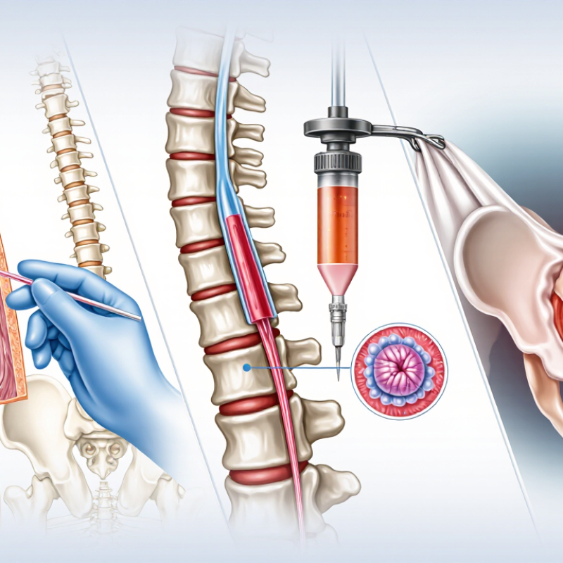
A core needle biopsy removes narrow cylinders of bone or soft tissue through a small incision. It often provides enough material for microscopic and molecular testing. An open biopsy uses a larger incision when needle sampling is inconclusive or difficult. Incisional biopsies remove part of a lesion, while excisional biopsies remove the entire abnormal area. Excision is less common for large or deep bone tumors. The biopsy route should be planned with the specialist who may later perform tumor surgery. Poor placement can complicate treatment.
The World Health Organization’s tumor classification emphasizes accurate tissue diagnosis because bone tumors can look similar on scans. Pathologists may test cell structure, proteins, or genetic changes. The process is not always tidy. A sample can be too small, damaged, or unrepresentative. That possibility deserves discussion before the procedure. Patients should ask who will perform the biopsy, how pain will be managed, and when results are expected. According to NCI guidance, specialist review can be valuable when the diagnosis is unusual or uncertain.
Fine-Needle Aspiration Versus Core Needle Biopsy: Cytology and Tissue Architecture
Fine-needle aspiration (FNA) uses a thin needle to collect loose cells from a bone lesion. Cytology can reveal malignant cells, infection, or a benign process quickly. It is less traumatic and may suit a fluid-filled or easily accessible lesion. However, FNA usually cannot show tissue architecture. That missing structure can make lymphoma, sarcoma, and some bone-forming tumors difficult to classify.
Core needle biopsy removes narrow tissue cylinders. Pathologists can examine cell arrangement, matrix production, necrosis, and invasion patterns. These details often guide tumor grading and treatment planning. A 2021 systematic review in European Radiology reported approximately 93% diagnostic yield for image-guided percutaneous bone biopsy, with major complications near 1%. The figure is useful, but it does not guarantee success for every lesion.
Imaging guides the choice. A radiologist may select FNA when cytology can answer a focused question. Core biopsy is commonly preferred when architecture affects diagnosis. The American College of Radiology’s bone tumor guidance stresses biopsy planning with the treating surgical team. Poorly placed tracks can complicate later surgery. That risk deserves attention.
The boundary is not perfectly clean. Some cases need both methods. Small samples can also mislead, especially after treatment or in heavily mineralized lesions. My practical concern is simple: a fast procedure is not always the most informative one. The biopsy method should match the suspected disease, lesion location, imaging pattern, and required pathology tests.
Open Surgical Biopsy: When Needle Sampling Is Inconclusive or Insufficient
Top Bone Biopsy Procedure Types: What You Need to Know?
Open surgical biopsy may be recommended when needle sampling is inconclusive or provides too little tissue. This procedure uses a small incision to expose the bone or nearby lesion. A surgeon removes part or all of the targeted area for laboratory examination. It is usually planned with an orthopedic surgeon, radiologist, and pathologist. Their coordination matters because the biopsy path can affect later treatment.
This approach may help when a lesion has mixed tissue types, lies deep within bone, or requires its overall structure for diagnosis. It can also clarify uncertain results from imaging or needle biopsy. However, open biopsy is more invasive. Possible concerns include bleeding, infection, nerve injury, anesthesia reactions, and a longer recovery. The exact risk depends on the lesion’s location and the patient’s health. No biopsy plan is perfect. Even experienced teams may need to reconsider the approach after new imaging or pathology findings.
Tips: Ask why open biopsy is preferred over another method. Confirm which specialist will perform it and how the sample will reach pathology. Discuss pain control, activity limits, wound care, and warning signs such as fever or increasing swelling. Bring previous scans and pathology reports. Small details can change the plan. Be honest about medicines, allergies, and past surgical problems.
Biopsy Safety: Major Complication Rates Are Typically Below 1% in Studies
Top Bone Biopsy Procedure Types: What You Need to Know
Bone biopsy safety deserves careful attention. The main options include image-guided core needle biopsy, fine-needle aspiration, and open surgical biopsy. A core needle removes a small tissue cylinder for detailed laboratory analysis. Fine-needle aspiration collects cells with a thinner needle. Open biopsy uses a larger incision and may be considered when needle sampling cannot provide enough tissue. The choice depends on the lesion’s size, location, appearance, and suspected diagnosis.
Studies commonly report major complication rates below 1% for bone biopsy in selected patients and experienced centers. “Major” usually means events requiring significant treatment, not every bruise or short-lived ache. Possible problems include bleeding, infection, fracture, nerve irritation, or a nondiagnostic sample. Risk may increase near the spine, pelvis, major blood vessels, or weight-bearing bones. The statistic is reassuring, but it is not a personal forecast.
Before the procedure, clinicians should review imaging, blood tests, medicines, allergies, and bleeding history. An experienced specialist should plan the safest needle path with the interpreting pathologist. Patients should understand who will monitor them and when results are expected. Clear aftercare matters. Increasing pain, fever, weakness, or persistent bleeding requires prompt medical advice. I would question any explanation that promises zero risk or treats one study as universal.
